Both halves of this claim are well supported, and neither rests on a single study. About 8 percent of the human genome, by the standard estimate, consists of sequences left behind by ancient retroviruses. And among those sequences are a small number of genes that the body now uses to build the placenta. The viral DNA is not a curiosity sitting inertly in the genome. Some of it does work.
This is a body of research assembled over roughly twenty-five years by several independent groups, not one paper that might not replicate. The broad outline is settled. The parts that remain genuinely uncertain are narrower, and worth separating out.
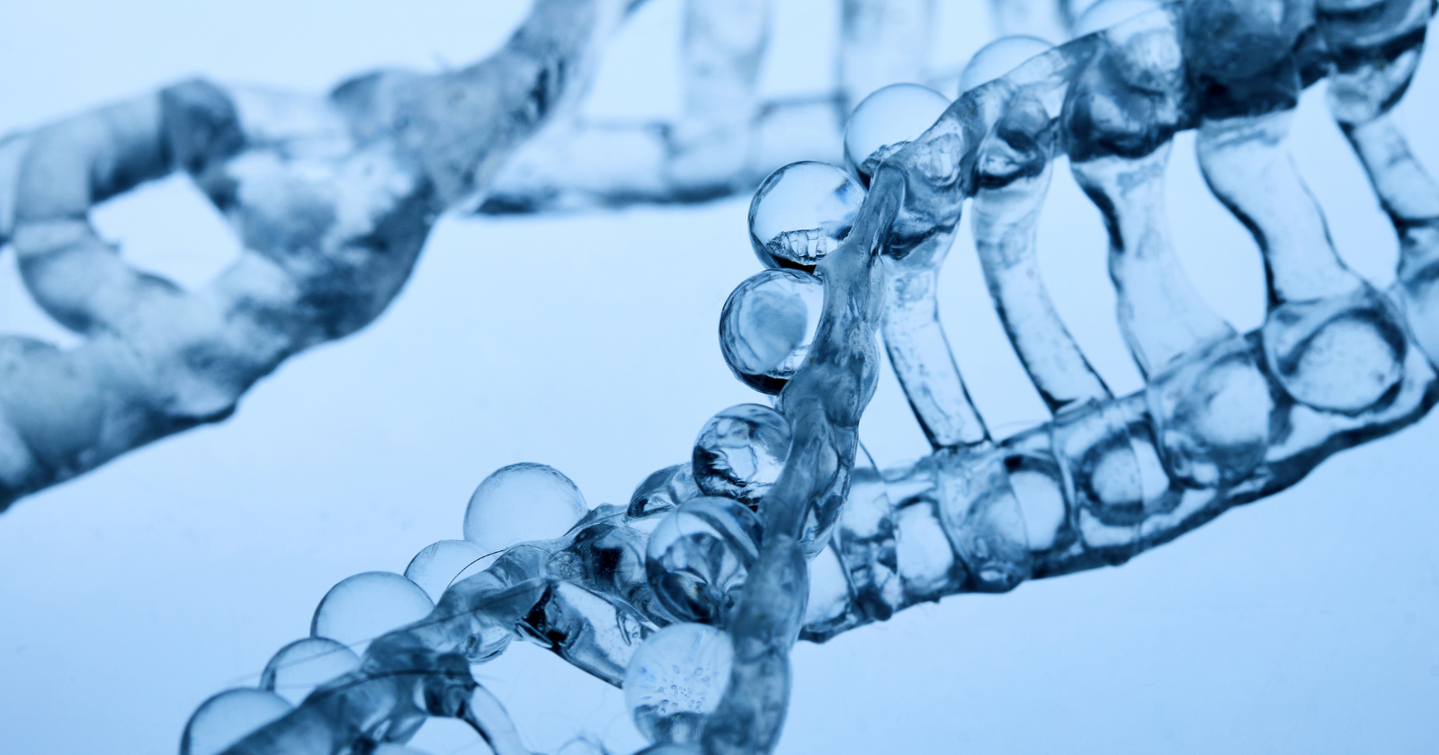
How viral DNA becomes permanent
Retroviruses copy themselves by inserting their genetic material into the DNA of the cells they infect. Usually those are body cells, and the inserted sequence dies with the host. Occasionally a retrovirus infects a germline cell, an egg or sperm or one of their precursors. When that happens, the viral sequence can be passed to offspring, and from them to their offspring, until it becomes a fixed part of the species’ genome.
Sequences acquired this way are called endogenous retroviruses. According to a 2022 review in Physiological Genomics, they make up around 8 percent of human DNA, more than four times the share occupied by protein-coding genes, and they accumulated across roughly the last 100 million years of primate and pre-primate evolution. Most of them are genomic fossils: mutated, fragmented, or silenced remnants that can no longer behave like infectious viruses. For a long time they were filed under the unhelpful heading of junk DNA.
A few are not fossils. A small number of these viral genes were kept, in working order, because they turned out to be useful. Biologists call this co-option, or exaptation: a part that evolved for one purpose put to another.
What the placenta borrowed
The clearest case is a group of genes called syncytins.
A retrovirus carries an envelope gene, known as env, that produces a protein on the virus’s surface. The protein’s job is to fuse the virus’s membrane with the membrane of a target cell, the step that lets the virus get inside. That fusion machinery is what was repurposed.
The human placenta has an outer layer, in direct contact with the mother’s tissue, called the syncytiotrophoblast. It is not made of separate cells. It is a single continuous structure containing many nuclei, formed when individual cells fuse together. The fusion is carried out by syncytin proteins, which are produced from co-opted retroviral env genes and have kept the membrane-fusing ability of their viral ancestor. The gene that does this in humans, syncytin-1, derives from a retrovirus designated HERV-W. It was identified in 2000 by Mi and colleagues in a paper in Nature describing a captured retroviral envelope protein active in placental development. Humans have a second such gene, syncytin-2, from a separate and older viral insertion.
Where the proof actually comes from
It is worth being precise about the word “help” in the claim, because the strongest evidence is not human.
You cannot delete a gene in a human to see what breaks. The decisive functional test was done in mice, which carry their own syncytins, syncytin-A and syncytin-B, captured independently from a different retrovirus. In a 2009 study in PNAS, Anne Dupressoir, Thierry Heidmann, and colleagues knocked out syncytin-A in mice. The placental fusion layer failed to form properly, maternal-fetal exchange was impaired, and the embryos died partway through gestation. That is direct evidence that a co-opted viral gene is not decorative but required.
For the human genes, the evidence is strong but inferential. In mice, where the decisive knockout experiments can be done, syncytin genes are necessary for normal placental formation and embryo survival. The human genes carry the same major signatures: placenta-specific expression, cell-fusion activity in laboratory assays, and conservation under purifying selection over tens of millions of years, which is the genome’s way of indicating a sequence is doing something it cannot afford to lose. “Help make pregnancy possible” is a fair summary, provided the reader understands that the human part of the claim is built on expression, function in cell culture, and conservation, supported by analogy with the animal knockouts, rather than on a knockout no one can ethically perform.
The same trick, captured again and again
The stranger finding is what turned up when researchers looked across other mammals.
Primates, mice, rabbits, carnivores, ruminants, and guinea pigs all have syncytins. But they are not the same gene inherited from a shared ancestor. They come from different retroviruses, captured separately in each lineage at different points in time. A 2013 paper in PLOS Genetics from Heidmann’s group describes these genes as necessary functions acquired by chance, repeatedly and independently. Even a live-bearing lizard, the Mabuya skink, has been found to have captured one.
So the placenta was not assembled once with a viral component bolted in. The same category of viral part has been recruited over and over, by separate lineages, for the same job. The Heidmann group has gone further and proposed that a single founding capture, perhaps 150 million years ago, was pivotal to the original emergence of placental reproduction, with later viral genes replacing the founder in different lineages. That last idea should be read as a hypothesis under active investigation, not a settled result. The independent captures are documented. The founding-event story is an interpretation laid over them.
It is also worth keeping the placenta case separate from the rest of the field. Endogenous retroviruses turn up in research on autoimmune and neurological disease, and some of that work is genuinely contested. The placental syncytins are the part of the story where the evidence is clear, and they do not carry the disease literature on their back.
What it changes
The picture of the genome as a tidy blueprint of human genes was always a simplification. A more accurate image is a sediment, with layers of old infection compacted into it, most of it inert. What the syncytin work shows is that the sediment is not uniformly dead. At least one layer of it is structural.
The open question is how much else is doing real work. Cataloguing which co-opted viral sequences are functional, and which are simply old debris that has not yet been cleared, is slow and still unfinished. The syncytins are the clearest answer so far. They are unlikely to be the only one.