

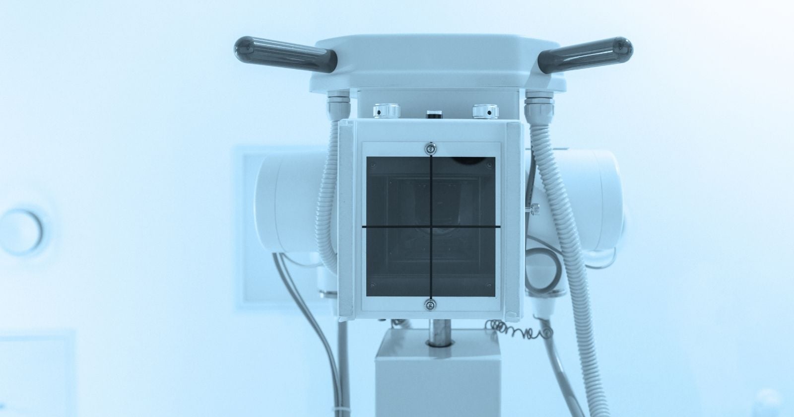
On March 31, 2025, four first-time astronauts flying the first human mission ever to loop directly over Earth’s poles carried a commercial off-the-shelf X-ray machine into orbit — a wireless MinXray unit smaller than a briefcase — and used it to take the first diagnostic radiographs ever produced in space. The images were of each other’s hands, forearms, chests, abdomens and pelvises, plus a phantom calibration target and a smartwatch. They were rated by independent radiologists as equivalent in quality to the same crew members’ preflight scans on Earth.
The experiment sounds modest. Its implications are not.
Every serious plan to send humans back to the Moon, and eventually to Mars, runs into the same wall: what happens when someone breaks a bone, develops a kidney stone, or takes a hard fall on a dusty slope hours from the lander. Earth-based emergency medicine leans heavily on imaging. Off-planet medicine, until Fram2, did not.
What Fram2 actually did
The Fram2 mission flew commander Chun Wang, vehicle commander Jannicke Mikkelsen, pilot Rabea Rogge and mission specialist Eric Philips on a 3-day, 14-hour flight aboard the SpaceX Crew Dragon Resilience. It was the first human spaceflight to fly a 90-degree orbit, sending the crew directly over the north and south poles at an altitude of roughly 425 to 450 kilometers.
Tucked into the mission’s science manifest was a compact wireless radiography system — an FDA-cleared MinXray Impact Wireless generator paired with a flat-panel digital detector. Three of the four crew received four hours of preflight training on the device. None of the four are physicians.
Once in orbit, the crew imaged each other and a piece of hardware — a smartwatch, chosen as a stand-in for the kind of equipment inspection that could matter on a long mission — with no live guidance from the ground. Independent radiologists then compared the space-based images against preflight X-rays taken on Earth. Their verdict, published this month in the RSNA journal Radiology: the in-flight radiographs were equivalent to preflight images in overall image quality, spatial resolution and contrast resolution. Positioning for chest, abdomen and pelvis views was harder in microgravity and scored somewhat lower, but every image was rated diagnostic.
The generator came home on the Dragon with only superficial damage to its housing. Its internal hardware and X-ray output were unaffected.
Why bones matter more the farther you go
Astronauts on the International Space Station lose bone density in weight-bearing regions of the skeleton during extended stays. Fracture risk climbs accordingly, particularly during the physical shock of re-entry or the first days back under gravity. A lunar surface mission adds falls on uneven regolith, EVA-suit trauma, and equipment strikes to the list. Returning astronauts already describe pain in the soles of the feet and lower back within days of splashdown, before anyone falls off anything.
The ISS has managed without diagnostic radiography because it can be evacuated. A Soyuz or Dragon can bring a sick crew member home in hours. That safety net does not stretch to the Moon, and it certainly does not stretch to Mars.
The point was made sharply in January, when NASA cut short its Crew-11 mission on the ISS after a crew member developed an undisclosed but “apparently serious” medical condition. The agency canceled a scheduled spacewalk on January 7 and, the next day, announced Crew-11 would come home more than a month early — the first medical evacuation in the station’s 25-year history. The Dragon splashed down off San Diego on January 15. NASA officials said the decision was made in part because the diagnostic tools they wanted for the patient existed on Earth, not aboard the station.
On a lunar surface mission, that option collapses. On a Mars transit, it disappears entirely.
The training question
Deep-space crews will be small. Two, three, maybe four people. Not everyone can be a physician, and even if one crew member is medically trained, that person could be the patient. On Fram2, mission specialist Eric Philips is a professional polar guide, not a doctor.
The Fram2 result matters partly because it addresses that specific problem. The experiment demonstrated more than simply that radiography works in microgravity — it showed that non-experts, trained for a single afternoon, could produce diagnostic-grade images without live coaching from a flight surgeon.
The team behind the experiment, led by Dr. Sheyna Gifford, a Mayo Clinic aerospace-medicine physician, first tested the device on a parabolic aircraft flight in 2022 — the so-called Vomit Comet — which simulates weightlessness in bursts of roughly 20 seconds. Fram2 was the leap from a physics demonstration to an operational one.
Hardware inspection, and a dual-use dividend
The smartwatch image is easy to overlook. It shouldn’t be.
Long-duration missions fail more often from equipment problems than from medical ones. Cracked circuit boards, degraded seals, fatigued welds — these are the kinds of faults that ground engineers currently diagnose by talking a crew through visual inspection or waiting for a component to arrive back on Earth. A portable X-ray gives a lunar base the ability to look inside a failing part without disassembling it, or to confirm that a printed replacement matches spec. The Fram2 images of the smartwatch resolved internal components at submillimeter scale.
Space medicine has a long history of producing technology that ends up helping people who will never leave Earth. Portable radiography developed for austere spaceflight environments plugs directly into the needs of rural clinics, disaster zones and battlefield medicine — places where a fixed radiography suite is unimaginable. The MinXray system on Fram2 is already the sort of unit paramedics use at the Kentucky Derby and on the sidelines of the Super Bowl, as Gifford has pointed out. Space is just the newest austere environment.
What this does not prove
A few caveats deserve to sit in the open.
Fram2 was a short-duration mission in low Earth orbit. Radiation exposure was modest, thermal cycling was benign, and the crew was healthy. A three-year Mars mission looks nothing like that. Detectors degrade in high-radiation environments. Consumables run out. Software fails.
The imaging sample was also small — seven anatomic radiographs, on healthy crew members, taken with no urgency. Diagnosing a clean tibial fracture is one thing. Reading a subtle pulmonary embolism on a chest X-ray taken by a crewmate with four hours of training is another. The Fram2 result is a proof of feasibility, not a proof of clinical parity.
And X-rays are only one imaging modality. Kidney stones — a recurring risk in microgravity due to calcium loss — are usually diagnosed with ultrasound or CT, not radiography. A lunar clinic will need more than one instrument.
The direction of travel
Still, the trajectory is clear. Commercial short-duration missions like Fram2 are increasingly being used as testbeds for the medical and operational hardware that Artemis and its successors will depend on. Artemis II already carried its own layered biomedical research program on the crew’s April 2026 lunar flyby. The private spaceflight sector is becoming, in effect, an applied research arm for lunar and Martian mission planning — testing the tools that government programs will later fly to the lunar south pole and beyond.
A portable X-ray in orbit is a small step. It is also the sort of quiet, unglamorous engineering advance that separates a flags-and-footprints mission from a sustained human presence off Earth. Sustained presence requires the ability to treat injuries in place, inspect hardware without shipping it home, and give small crews real diagnostic tools instead of a satellite call to a flight surgeon and a hope for the best.
Chun Wang and his crew were in orbit for three and a half days. The little wireless generator they carried up with them, imaged each other with, and brought back down mostly intact, may end up mattering for decades.