
Albert Einstein died on April 18, 1955, in Princeton, New Jersey, at the age of 76. The cause of death was an abdominal aortic aneurysm. The autopsy was performed at Princeton Hospital by the pathologist on duty, a Yale-educated physician named Thomas Stoltz Harvey. Einstein had requested that his body be cremated and his ashes scattered without ceremony, to discourage the kind of posthumous cult he had spent decades trying to avoid in life. The cremation occurred. The scattering occurred. What did not occur was the cremation of the entire body.
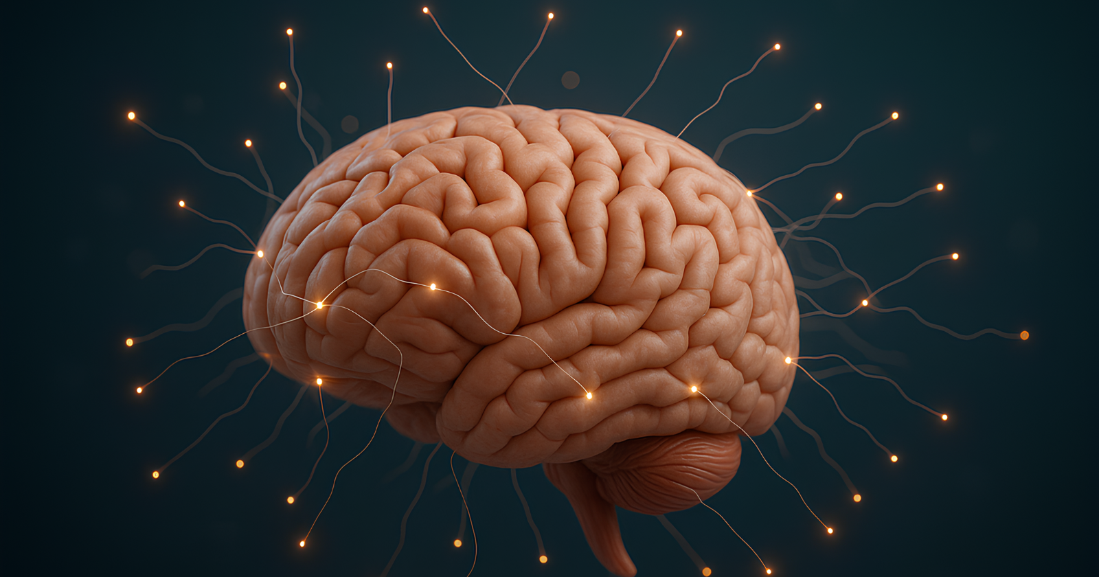
Harvey had, during the autopsy, removed Einstein’s brain. The removal had not been authorized by Einstein’s family. It had not been authorized by Einstein himself. It was, by any honest accounting, an act of medical theft. Harvey would, in the immediate aftermath, retroactively secure permission from Einstein’s elder son Hans Albert, on the condition that the brain would be used only for legitimate scientific research. The permission was unenforceable. What followed was a series of events that medicine has never fully come to terms with.
What Harvey did with the brain
Harvey photographed the brain from multiple angles, weighed it, measured it, and dissected it into approximately 240 separate sections. He preserved the sections in formaldehyde and other fixatives. He prepared microscope slides from selected portions and sent them to a small number of neuroscientists he considered qualified to study them. He kept the rest for himself.
The keeping went on for forty years. Harvey moved frequently across the decades following Einstein’s death. He lost his position at Princeton Hospital, in part because of his refusal to return the brain. He drifted through small towns in the American Midwest. He divorced. He worked, for a period, in a plastics factory in Wichita, Kansas. The brain traveled with him. For long stretches, it was stored in two mason jars inside a cardboard box that had originally contained Costa Cider. The University of Washington’s documentation of this period notes that a journalist named Steven Levy, writing for New Jersey Monthly in 1978, tracked Harvey down in Wichita and found the brain in exactly this configuration. The box sat in Harvey’s office, alongside the ordinary objects of a man’s working life.
Most of the scientific community, throughout these decades, had assumed the brain was lost. It was not lost. It was sitting in a cider box in Kansas, waiting for someone to ask the right questions.
What Marian Diamond found
The first substantial scientific study emerged in 1985, thirty years after Einstein’s death. It was led by Marian Diamond, a neuroscientist at the University of California, Berkeley, working with collaborators including Arnold Scheibel, Greer Murphy, and Harvey himself, who was listed as a co-author. The Cold Spring Harbor Laboratory’s documentation of the study describes the methodology. The team had access to sections from four specific regions of the brain: Brodmann area 9 in the prefrontal cortex and Brodmann area 39 in the parietal lobe, on both hemispheres.
They counted the cells. Specifically, they counted the ratio of glial cells to neurons in each of the four regions. Glial cells are the support cells of the brain, providing structural and metabolic assistance to the neurons that conduct the actual signaling. The ratio is one indicator of how metabolically demanding the surrounding neural tissue is.
In the left area 39 of Einstein’s brain, the team found a glial-to-neuron ratio significantly higher than what they measured in eleven control brains. The other three regions did not show similarly significant differences. The implication was that the neurons in this part of Einstein’s parietal lobe had been metabolically more active than the corresponding neurons in ordinary brains.
The study was published in Experimental Neurology. It was also more cautious than its popular retelling has tended to suggest. The sample size was small. The control brains had not been matched to Einstein in age, sex, or other demographic features. The methodology had limits the authors themselves acknowledged in the paper. The result was suggestive rather than conclusive. It was absorbed as conclusive anyway.
What Sandra Witelson found
The more substantial finding came fourteen years later, in 1999, when the Canadian neuroscientist Sandra Witelson at McMaster University in Hamilton, Ontario, published a study in The Lancet that examined the overall anatomy of Einstein’s brain using the photographs Harvey had taken in 1955.
Witelson worked from the original photographs, taken before the brain was sectioned, and from a small number of preserved brain blocks. She compared the dimensions of Einstein’s brain to a control sample of 35 normal male brains from her own institution’s brain bank. The published account of her analysis describes the central finding. The inferior parietal lobule in Einstein’s brain was approximately 15 percent wider than the same region in the control sample. The widening was visible in both hemispheres, and substantial by the standards of how brain anatomy normally varies.
The inferior parietal lobule is the region most consistently associated with mathematical reasoning, spatial cognition, and the visualization of three-dimensional relationships. The association is not incidental to Einstein’s intellectual work. He had described his own thinking process, on multiple occasions, in terms that emphasized visual and spatial imagery rather than verbal reasoning. He famously conducted what he called “thought experiments,” in which he imagined himself riding alongside beams of light, falling alongside elevators, and running other spatial visualizations that physicists have, in the decades since, recognized as central to his most important contributions.
Witelson’s study also identified a second anomaly. The brain partially lacked the Sylvian fissure, or lateral sulcus, the prominent groove that normally separates the parietal and temporal lobes. In Einstein’s brain, the fissure was foreshortened or partially absent in both hemispheres. The implication, on Witelson’s analysis, was that the inferior parietal lobule was not only larger than average but also more integrated with the surrounding tissue than the standard anatomical configuration would allow.
What the headlines tended to skip
The honest acknowledgment is that the findings about Einstein’s brain are far less conclusive than their popular framing makes them sound.
Every study so far has been conducted on a single brain, with control samples that have varied in quality and matching. None has established, statistically, that the anatomical features being identified are causally connected to Einstein’s intellectual capacities. The features could be coincidental. They could equally be the product of his decades of mathematical and physical thinking, rather than the cause of it. The available methodology has not been able to distinguish between the two possibilities.
A 2012 study by the anthropologist Dean Falk, working with additional photographs Harvey had taken that had not previously been examined, identified further anomalies, including an unusual knob-like structure on the motor cortex often observed in left-handed musicians, which Einstein was. The NPR documentation of Falk’s work describes her additional finding that Einstein’s corpus callosum, the bundle of fibers connecting the two hemispheres, was thicker than normal, suggesting greater communication between the hemispheres than the standard configuration would produce.
All of these findings are interesting. None definitively establishes the causal relationship between Einstein’s anatomy and his cognition that the headlines have wanted. Witelson herself was explicit about this in her 1999 paper, noting that her work “clearly does not resolve the long-standing issue of the neuroanatomical substrate of intelligence.” The headlines absorbed the 15 percent finding without absorbing the caveat.
Final words
Einstein’s brain was removed from his body during the autopsy on April 18, 1955, by a pathologist who did not have the family’s permission to remove it. The brain was kept in a series of jars across the next four decades, traveling with the pathologist as he moved from Princeton to small towns in the American Midwest, eventually ending up in a cardboard box marked “Costa Cider” in an office in Wichita, Kansas. Harvey lost his career, his marriage, and his standing in the scientific community over his refusal to give it back. He spent long stretches of his working life in a plastics factory. He died in 2007.
The brain itself, once its continued existence was confirmed, became the subject of a small number of scientific studies that identified various anatomical anomalies. The most widely cited, from Witelson’s 1999 study, was that the inferior parietal lobule was about 15 percent wider than the same region in 35 control brains. That part of the brain is the one most consistently associated with the mathematical and spatial reasoning Einstein’s work depended on.
The findings are less conclusive than they are popularly treated as. They rest on a single brain. None has shown that the features being identified caused the cognition they appear to correlate with. They are suggestive, not definitive. The clean answer the story has wanted is not one the underlying evidence can provide. What the studies actually established is worth reading on its own terms, before reaching for the conclusions about genius and anatomy that the headline summaries have been built around.
