
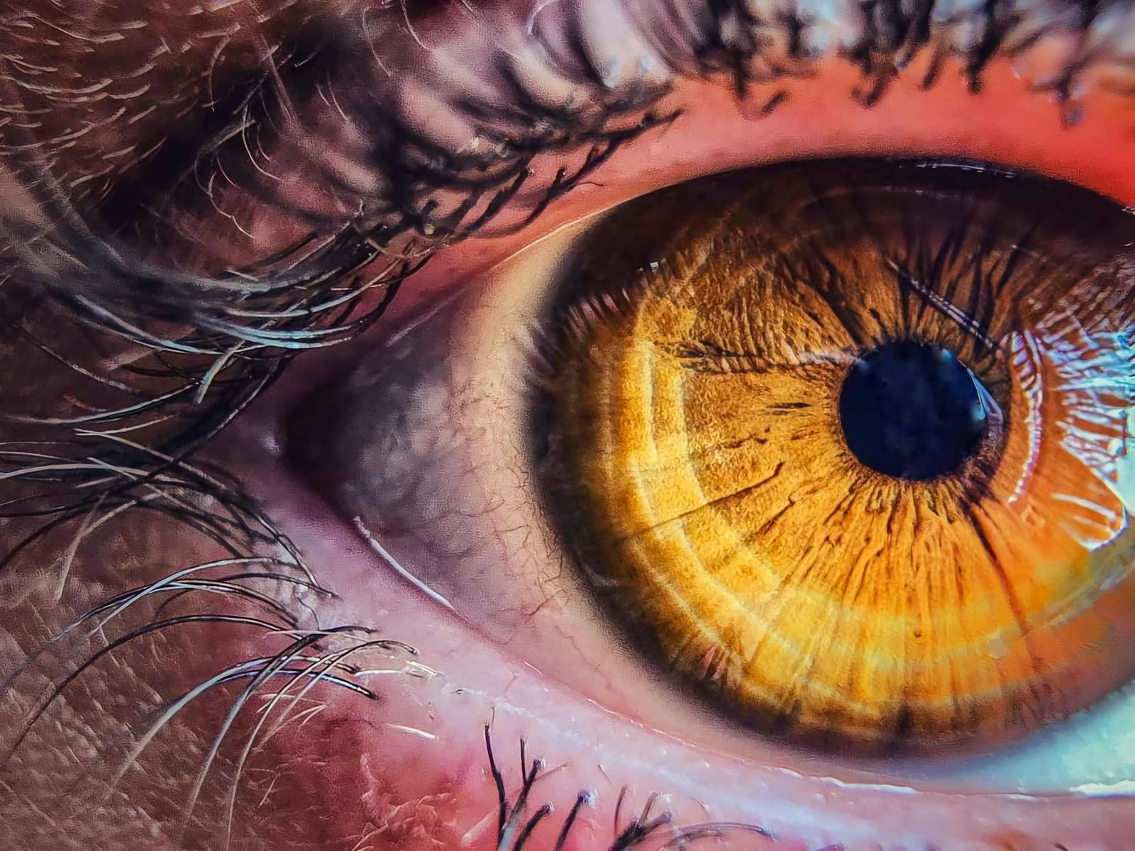
The photoreceptors at the back of your eyes are neurons. Not eye cells in some specialised sense, but actual brain tissue, the same lineage of cells that builds your cortex and your spinal cord, pushed outward through the eye stalk during early embryonic development until they reach the surface of the skull and meet the world. When light from Betelgeuse, a distant star hundreds of light-years away, strikes your retina on a clear night, it is striking neural tissue that started its life inside the developing forebrain. The photons travelled for centuries to land on a piece of your brain.
This is not metaphor. It is embryology.
The eye is an outpouching of the brain
Early in human gestation, the neural tube, the structure that will become the entire central nervous system, develops two small bulges near what will become the front of the brain. These are the optic vesicles. They balloon outward, contact the overlying ectoderm, and then invaginate to form a two-layered cup. The inner layer of that cup becomes the neural retina. The outer layer becomes the retinal pigment epithelium. The stalk connecting it all back to the brain becomes the optic nerve. The National Library of Medicine’s Webvision reference puts the relationship plainly: the retina is a part of the central nervous system and derives from the neural tube.
That optic nerve is not a nerve in the peripheral sense. It is a tract of the central nervous system, sheathed in CNS myelin. Cut it and it does not regenerate, because CNS tissue does not regenerate the way peripheral nerves do. The retina is brain. The optic nerve is brain. The light-sensing surface at the back of your eye is a piece of cortex that got displaced during the first month of your existence and never came home.
Researchers at Johns Hopkins recently mapped one of the most delicate parts of this process. In a study published in Proceedings of the National Academy of Sciences, biologist Robert J. Johnston Jr. and his team used lab-grown retinal organoids to show that a vitamin A derivative called retinoic acid and thyroid hormones coordinate to pattern the foveola, the tiny central region of the retina critical to human visual perception. During fetal development, blue cones in that central patch convert into red and green cones. The eye is still rearranging its photoreceptors months before birth.
Why this lineage matters for what you see
The reason photoreceptors are neurons, and not some specialised epithelial cell, is that they need to do something only neurons can do well, which is fire signals to other neurons with sub-millisecond timing. A rod cell responds to individual photons. It does this by changing the shape of a molecule called rhodopsin, which triggers a cascade that closes ion channels in the cell membrane and changes the cell’s electrical potential. That signal then passes through bipolar cells, amacrine cells, and ganglion cells, all of them also neurons, all of them part of the same neural tissue that grew out of the forebrain.
By the time a signal leaves your eye through the optic nerve, it has already been processed by three layers of brain. The retina is not a camera sensor sending raw data inward. It is a piece of cortex doing edge detection, motion detection, and contrast normalisation before anything reaches the visual centres at the back of your skull. The progenitor cells that build your retina are the same kind of cells that build your hippocampus. They took a different turn during gestation, but they began in the same place.
The photon’s journey to your brain
Consider what happens when you stand outside on a clear night and look at Vega, the bright star in the summer constellation Lyra, about 25 light-years away. A photon left the surface of that star around the year 2001. It travelled across roughly 237 trillion kilometres of mostly empty space. It entered the upper atmosphere, was refracted slightly by air of varying density, passed through your cornea, your aqueous humour, your lens, your vitreous gel, and finally struck a cone cell at the back of your eye.
That cone cell is a neuron whose cell body sits in the outer nuclear layer of your retina, with its light-sensing outer segment pointing back toward the pigment epithelium. The photon is absorbed by an opsin molecule embedded in stacked membrane discs. The opsin’s retinal cofactor, a derivative of vitamin A, isomerises from 11-cis to all-trans. A G-protein cascade fires. The cell hyperpolarises. A signal moves inward.
For starlight from Vega, that means the photon ended its 25-year journey on a piece of tissue that is, anatomically and embryologically, part of your central nervous system. Your brain caught it.

An evolutionary inheritance much older than vertebrates
The neural origin of photoreceptors is not a human quirk. It is ancient. In every jawed vertebrate, in fish and reptiles and birds and mammals, the retina forms in the same way: as an outpouching of the embryonic forebrain that meets the surface ectoderm, invaginates, and becomes a layered tissue of neurons. Even in lampreys and hagfish, the jawless fish whose ancestors diverged from the rest of vertebrates deep in evolutionary time, the eye is built from the same neural-tube evagination that builds yours.
The pattern is older than jaws. Older than bones. Older than land.
What this means for medicine
The brain-tissue identity of the retina is not just a curiosity. It is the reason vision loss is so difficult to reverse. When photoreceptors die, in macular degeneration or in inherited retinal degenerations, the cells do not grow back. Brain neurons do not regenerate. This is why gene therapy and cell replacement have become the major fronts in retinal medicine.
At the University of Pennsylvania’s School of Veterinary Medicine, Raghavi Sudharsan and William A. Beltran have developed four novel photoreceptor-specific promoters for gene therapy at advanced disease stages, short DNA sequences under 850 base pairs that can be packaged into adeno-associated viruses and switched on inside surviving rods and cones even after half the retina has died. The work, published in Molecular Therapy, attacks the problem that most existing gene therapies were tested only in healthy eyes and lose effectiveness as the tissue degenerates.
A parallel approach is to grow new photoreceptors from scratch. A team at Wenzhou Medical University identified a population of human neural retinal stem-like cells capable of regenerating retinal tissue and restoring some vision in mice. Other labs are working on the developmental biology side, with researchers reporting that a specific enzyme helps maintain retinal progenitor cells during eye development, a finding that could improve the yield of usable cells in lab-grown organoids.
All of these efforts are, in effect, neurosurgery. They are attempts to repair, replace, or rewire neurons in a piece of the brain that happens to sit at the front of the skull.
The dark adaptation no one explains
If you walk from a brightly lit room into a dark one, your vision improves over the next twenty to thirty minutes. This is not your pupil dilating. The pupil finishes that job in seconds. What is happening over those long minutes is a chemical process inside your photoreceptor neurons, the regeneration of rhodopsin from its bleached state. The retinal cofactor has to be re-isomerised and recombined with the opsin protein. The pigment epithelium does most of this chemistry, shuttling vitamin A derivatives back and forth.
This is why looking at the night sky takes patience. Your brain, the part of it sitting at the back of your eye, needs time to ready itself. The faint smear of the Milky Way is not visible until those neurons have finished their molecular maintenance.
The strange intimacy of starlight
Most of the photons that hit your retina on a given night come from streetlamps, screens, and the moon. But on a dark site, away from cities, your eyes catch light from sources that are genuinely far away. Sirius, the brightest star in the night sky, is about 8.6 light-years off. The photons reaching you tonight left in 2017. The light from the Andromeda galaxy, the most distant object visible to the unaided human eye, left its source roughly 2.5 million years ago, when early Homo was just starting to make stone tools in East Africa.
Those photons travelled across cosmic distances, missed everything else, and ended their existence by being absorbed by an opsin molecule inside a neuron that is part of your brain. The chain of events that began with a fusion reaction in a stellar core ends with a chemical change in a piece of central nervous system tissue sitting behind your eyelid.
There is no other organ that does this. Your skin does not receive photons in any meaningful neural sense. Your ears receive pressure waves that have travelled, at most, a few kilometres. Your nose receives molecules that drifted across a room. Only your eyes, and specifically the brain tissue inside them, regularly interact directly with matter and energy that originated outside the solar system.
A coda on what is touching what
Strictly speaking, the photon does not survive the encounter. It is absorbed. Its energy is transferred to an electron in the retinal molecule, which changes the molecule’s shape, which sets off the rest of the cascade. So the brain is not touching the photon so much as catching it, the way a glove catches a ball that no longer exists once it has been caught.
The retinal molecule, the opsin, the membrane disc, the cone cell, the bipolar cell, the ganglion cell, the optic nerve fibre, all the way back into the lateral geniculate nucleus and the visual cortex, are one continuous piece of neural tissue that began as a small bulge on the side of a four-week-old embryo’s forebrain. That bulge grew outward, met the surface, and stayed there, waiting for light.
Now it is dark outside, and somewhere in your peripheral vision, a photon that left a star before any human alive was born is about to finish its journey on a piece of your brain.