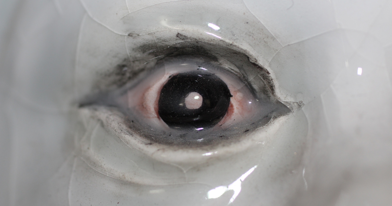
René Descartes decided the soul entered the body through the pineal gland, a lump of tissue roughly the size of a rice grain, buried near the centre of the brain. Palaeontologist Julien Benoit, writing in The Conversation, describes the same gland as a leftover: the shrunken remains of an organ that once sat on top of the skull and stared straight up at the sky.
That organ has a proper name, and a surprising number of animals alive today still have a working one.
A one-eyed filter feeder at the root of the tree
A review in Current Biology by George Kafetzis, Michael Bok, Tom Baden, and Dan-Eric Nilsson, working across the University of Sussex and Lund University, traces that relic back further than any fossil on its own could show. Their reconstruction has every vertebrate descending from a small, worm-like creature that lived in the ocean almost 600 million years ago, stayed put for most of its life, and fed by filtering plankton out of the seawater.
By then, the team argues, it had already lost the paired eyes on the sides of its head. Sitting still for a living makes side eyes expensive and largely useless. What survived was a patch of light-sensitive cells running down the middle of its head, which organised itself into one simple eye aimed upwards. A cyclops, sitting at the base of every backboned animal on the planet.
Later, when the lineage took up moving again, that central organ supplied the parts for a fresh set of paired eyes. Nilsson, professor emeritus in sensory biology at Lund, said the findings turn the accepted story of eye and brain evolution “upside down”.
Nobody has dug up the cyclops. Kafetzis and colleagues built their case by comparing where light-sensing cells sit and what they do across 36 major animal groups, then mapping that pattern onto the family tree. Soft tissue from the Precambrian almost never survives. The sequence is inferred, and the paper presents it as an argument rather than a settled result.
Why the vertebrate retina is the odd one out
Animal eyes draw on two ancient families of light-detecting cell, named ciliary and rhabdomeric after the structures they use to trap photons. In insects, squid and most other animals, rhabdomeric cells do the seeing in the side eyes while ciliary cells sit in unpigmented patches along the midline, handling background jobs such as tracking how bright the day is.
Human retinas run both at once. Rods and cones come from the ciliary branch. Ganglion, amacrine and horizontal cells, the layers that process a signal before it heads for the brain, trace back to the rhabdomeric one. That combination is a vertebrate peculiarity, and the authors trace it to a central eye that already carried both kinds of cell.
Animals that still have the spare
Tuatara have one. So do most lizards, many frogs and salamanders, lampreys, and a handful of sharks and bony fish. Properly called the parietal eye, it sits under a translucent scale on the crown of the head and comes complete with a lens and a stripped-back retina. In some frogs and lizards it shows up as a pale spot between the ordinary eyes. Images are beyond it. Light levels are not, and that alone tells an animal how the day length is shifting, when to bask, and when to breed.
The hole in the skull that closed
Bone keeps the receipts. A parietal eye needs a tunnel through the skull roof for its nerve, the pineal foramen, and that tunnel is obvious in a fossil. Benoit and colleagues at the University of the Witwatersrand examined more than 600 skulls from South Africa’s Karoo deposits, spanning 300 to 200 million years ago, in a paper published in Acta Palaeontologica Polonica.
Before roughly 260 million years ago, most pre-mammalian therapsids had the opening. After that it shrank and disappeared again and again in separate lineages, and among the cynodonts, the branch that eventually produced mammals, the foramen sealed over for good around 246 million years ago. The reading Benoit offers is that a third eye became dispensable once warm-bloodedness took over the job of holding body temperature steady, a scenario he says needs more testing.
An organ still running on light, secondhand
Nothing reaches the human pineal gland directly any more. Light lands on melanopsin-carrying ganglion cells in the retina, which pass word along the retinohypothalamic tract to the suprachiasmatic nucleus, the master clock tucked into the hypothalamus. From there the message detours out through the sympathetic nervous system and a relay in the neck before arriving at the pineal, which answers by producing melatonin in the dark. Fernanda Gaspar do Amaral and José Cipolla-Neto set out the whole circuit in Archives of Endocrinology and Metabolism.
Quite a detour for a gland that once did the job by looking up.
At 3am after a long flight east, the thing holding a person awake and staring at the ceiling is an eye that stopped seeing daylight about a quarter of a billion years ago, and has been taking directions through the post ever since.