

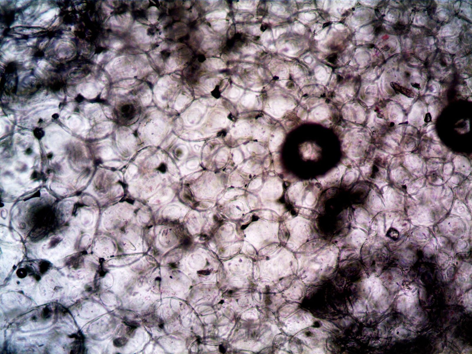
Push pieces of a living red encrusting sponge through a fine mesh and the animal disappears into a cloudy suspension of cells. Thousands of tiny bodies settle across the bottom of the dish, separated from the canals, chambers and surfaces that made them part of a sponge. Within minutes, some begin extending probing projections and moving towards their neighbours. The scattered cells start gathering themselves back into an animal.
Over the following hours, small clusters merge into larger masses. Over several days, some of those masses rebuild an outer surface, internal chambers and openings through which water can flow again. The result is not a repaired version of the body that entered the mesh, but a newly assembled sponge made from many of its surviving cells. A review of more than a century of sponge reaggregation experiments describes the process as one of the most dramatic examples of anatomical and cellular plasticity in animals.
What H. V. Wilson actually did in 1907
The experiment dates to the work of H. V. Wilson, an American zoologist at the University of North Carolina who conducted sponge research at the federal fisheries laboratory in Beaufort. Wilson had already observed that stressed sponges could simplify their tissues and later regenerate. In 1907, he took the process further by deliberately breaking sponge tissue into its constituent cells.
Wilson worked principally with Microciona prolifera, a sponge that begins life as an encrusting form and can later grow upright lobes. As he explained in his original paper on coalescence and regeneration, he cut the lobes into small pieces, folded fine bolting cloth around them and submerged the bundle in filtered seawater. He then squeezed it with forceps. The pressure broke apart the soft tissue while the cells passed through the cloth as red clouds.
Under the microscope, Wilson found several kinds of cells in the suspension. Granular amoeboid cells were especially conspicuous, but partially transformed collar cells and other pale cells were also present. The granular cells began collecting almost immediately, and the developing masses gradually incorporated other cells around them. Free cells and tiny clusters continued joining until larger, irregular aggregates covered parts of the glass.
Those aggregates did more than stick together. When Wilson spread them across slides and kept them in seawater, they flattened into encrusting masses and began differentiating. An outer membrane appeared, followed by connective material, flagellated chambers, canals and raised openings called oscula. In laboratory aquaria, Wilson reported that this degree of organisation developed in six or seven days, with active water currents visible in the reconstructed sponges.
From a red cloud to a working filter feeder
A sponge body is organised around water. Pores admit seawater, canals carry it through the animal and choanocytes use beating flagella to drive the current while trapping food particles. Water eventually leaves through one or more larger openings. Destroy that arrangement and the sponge temporarily loses the structure that allows it to feed and exchange gases efficiently.
Reaggregation begins before any new canal exists. Dissociated cells move with amoeboid motions, extending pseudopodia and thin contacts towards nearby cells. Small groups form first, then collide or draw together into larger aggregates. Modern live imaging of the Arctic sponge Halisarca dujardinii has recorded single cells attaching through slender projections during the first hours after dissociation.
Aggregation alone is not enough. Cells must change position, rebuild boundaries and restore the correct arrangement of feeding chambers and water channels. Surface-forming cells move towards the exterior, while other populations contribute to the internal structures. The smooth clump gradually acquires an inside, an outside and a route through which water can travel.
Success depends on species, temperature, seawater chemistry, cell density and the condition of the original tissue. Some aggregates remain simple clumps or die before completing the process. A study of the developmental checkpoints in sponge-cell aggregation found that the transition from loose cells to a functioning body proceeds through distinct stages, each of which can fail. Not every sponge species can complete the entire sequence.
How separated cells recognise the right partners
Wilson’s most revealing experiments involved mixing cells from different sponge species. He combined dissociated cells of Microciona with cells from Lissodendoryx, and in separate experiments mixed Microciona with Stylotella. Because the species had visibly different colours, he could follow their behaviour under the microscope. Instead of forming permanently mixed sponges, cells and developing masses preferentially joined members of their own species.
The result was a dish containing separate, species-specific aggregates. Microciona cells formed Microciona masses, while the other cells collected into their own groups. Some cells came into contact with the wrong species, but stable organisation occurred mainly among compatible cells. The experiment provided an early visual demonstration that animal cells can discriminate between different biological surfaces.
Later researchers identified extracellular adhesion and recognition systems, commonly discussed as sponge aggregation factors, that help mediate these contacts. Their exact composition and importance vary across sponge groups. The process is therefore more controlled than a simple tendency for any two sticky cells to collide and remain together.
Recognition solves only the first part of the problem. Compatible cells must still adopt the correct positions and produce the right proportions of different cell types. A functioning sponge cannot be built entirely from feeding cells or entirely from surface cells. Reaggregation is a sequence of recognition, migration, sorting, differentiation and tissue construction.
Stem cells help rebuild what the mesh erased
Archaeocytes play an important role in many sponges. These mobile, stem-like cells live in the mesohyl, the material between the animal’s outer surface and its internal feeding structures. They can divide and generate several specialised cell types. However, modern research no longer treats archaeocytes as the sole explanation for every sponge’s regenerative ability.
In a 1980 experiment, researchers isolated archaeocytes from the freshwater sponge Ephydatia fluviatilis and followed their development with electron microscopy. The purified archaeocyte aggregates began producing an outer pinacoderm after about 12 hours. Choanocytes appeared after 24 hours, followed by other specialised cells, demonstrating that archaeocytes from an adult sponge could retain extensive differentiation capacity.
Choanocytes can also contribute to sponge stem-cell systems. A review of sponge reproduction, regeneration and tissue maintenance describes archaeocytes and choanocytes as two major sources of cellular plasticity, with their roles varying among sponge lineages. Differentiated cells may also change identity through transdifferentiation. The rebuilding process draws on several kinds of cellular flexibility rather than one permanently pluripotent cell doing all the work.
A 2026 study of Halisarca dujardinii revealed another layer of control. Researchers detected active proteasomes at cell contacts during early reaggregation and found that inhibiting proteasome activity disrupted the development of aggregates. Their results indicate that sponge cells rapidly reorganise existing cytoskeletal machinery and protein-control systems as they migrate, attach and construct a new body.
Sponges are not the only animals that reassemble
The old claim that no other animal can regenerate from separated cells is not correct. Sponges remain the classic example, but related forms of reaggregation have been demonstrated in cnidarians. The differences concern the source of the cells, the reliability of the process and the developmental route taken afterwards.
In 1972, A. Gierer and colleagues reported that separated hydra cells reaggregated and developed into normal animals. Their experiment showed that hydra did not always require an intact tissue fragment to restore a body. Space Daily has also examined how the freshwater hydra continuously replaces its cells while preserving the same simple body plan.
Sea anemones provide another comparison. In 2026, researchers working with embryonic cells from Nematostella vectensis showed that reaggregated cells could restore their tissue layers and body axis, eventually producing whole polyps. The team found that a feedback relationship between Notch and Wnt signalling helped the disordered aggregate establish a single organised axis.
Those examples do not make the sponge experiment ordinary. Wilson began with cells from an adult animal, removed the visible architecture and obtained a new filter-feeding body with canals and flagellated chambers. More than a century later, sponges remain unusually tractable models of adult cellular plasticity. They are an extreme case, not an exclusive one.
What biologists can see between the mesh and the sponge
Reaggregation gives developmental biologists an unusual experimental starting point. Instead of watching an embryo build a body for the first time, researchers can erase much of an adult animal’s organisation and observe how its surviving cells reconstruct it. The dish exposes processes that are normally hidden inside intact tissue: recognition, adhesion, movement, sorting and differentiation.
Studies of ordinary sponge wounds reveal related molecular activity. Researchers examining injured Aplysina aerophoba found clotting-like responses, calcium signalling and changes involving Wnt and MAPK pathways during the hours and days after damage. A wound is not the same as complete dissociation, but both situations require cells to recognise disruption and restore tissue integrity.
Sponges accomplish this without neurons directing the work. That makes them relevant to the wider investigation of how the earliest animals coordinated specialised cells before conventional nervous systems evolved. Related research on tiny marine animals and the cellular origins of neurons shows how researchers are using simple living lineages to reconstruct the steps between local chemical communication and organised animal behaviour.
In the microscope dish, the reconstruction begins without a surviving canal to guide it. A cell stretches out a narrow projection, makes contact and joins a growing cluster; the cluster rounds, spreads and develops a boundary. Days later, flagella beat inside newly formed chambers and seawater moves through an opening that did not exist in the red cloud. The animal has not repaired its old architecture. Its cells have built another one.