

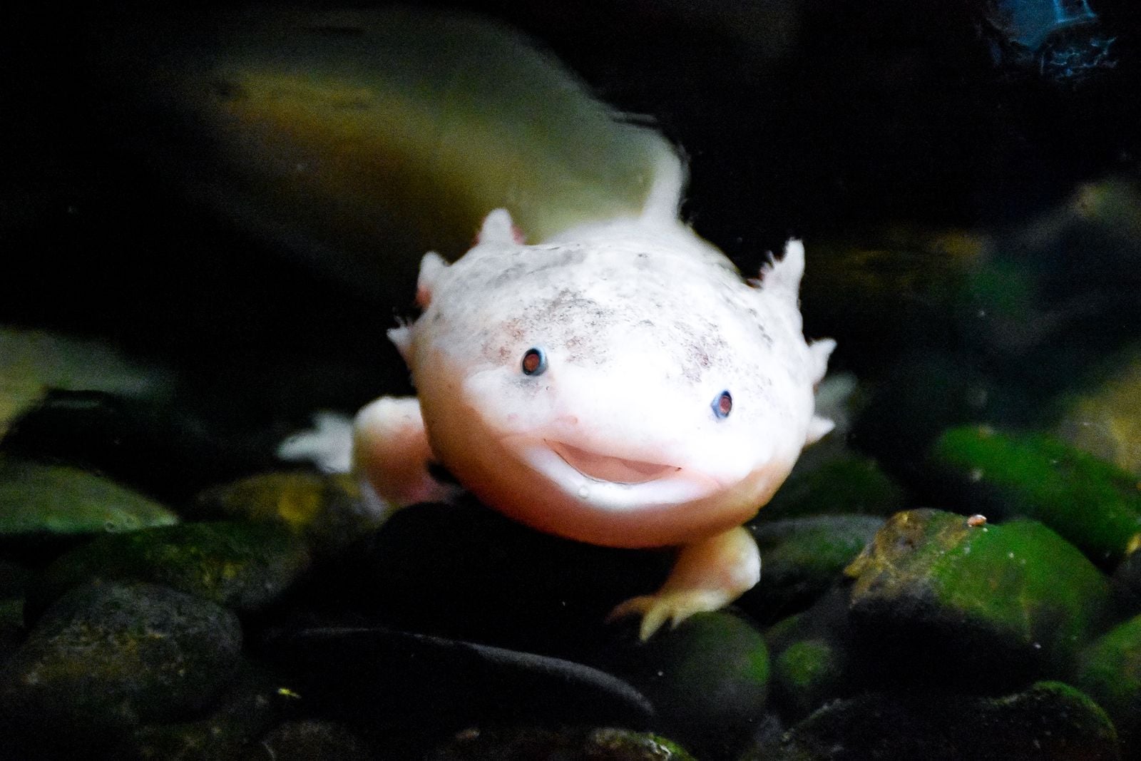
An axolotl can lose a leg and, given the right conditions, regrow it over several weeks to months — nerves, bone, cartilage, skin, blood vessels, five perfectly patterned toes, and no scar tissue where the wound used to be. The same salamander can regrow parts of its heart, sections of its spinal cord, chunks of retina, and even portions of its forebrain, producing brand new neurons that wire themselves into the surviving circuitry. In the wild, this animal exists in a single place: a shrinking network of canals and lake remnants at Xochimilco, on the southern edge of Mexico City, where the most recent surveys estimate fewer than a thousand adults remain.
The mismatch is almost hard to hold in one thought. An organism that has quietly solved problems human medicine has been circling for a century is being pushed out of existence by carp, tilapia, wastewater and urban sprawl in a lake system that was already ancient when the Aztecs built causeways across it.
What the axolotl can actually regrow
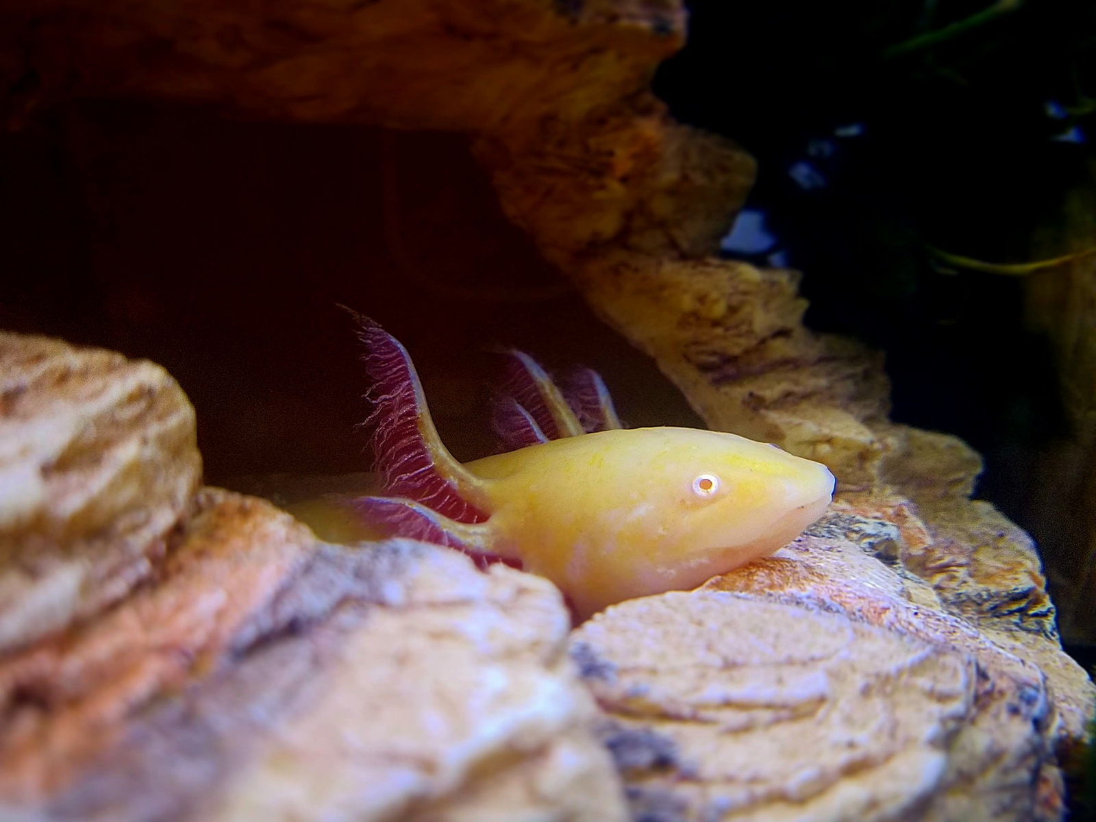
Ambystoma mexicanum is a neotenic salamander, which is a technical way of saying it never grows up. It keeps its feathery external gills, its finned tail and its aquatic life all the way through sexual maturity. It also keeps something more useful: a set of developmental programs most vertebrates switch off after embryogenesis.
The list of tissues it can rebuild is unusually long. Whole limbs, from shoulder or hip down. Jaws. Tail. Ovary and lung tissue. Portions of the heart. Segments of spinal cord, with function restored. Optic nerve fibres. And, as researchers have described, sections of the telencephalon — the forebrain region involved in sensory processing and behaviour — including newly generated neurons that migrate to the correct location and extend axons into the surrounding tissue.
The regenerated brain region ends up structurally similar to the original. Not a patch. A replacement.
How the trick works, and how the limb knows what to grow
When a limb is severed, the axolotl does something a mammal cannot. Instead of sealing the wound with a thick collagen scar, epidermal cells slide over the stump within hours to form a thin covering called the wound epithelium, which thickens into what biologists call the apical epithelial cap. Under this cap, cells beneath the injury begin to lose their adult identities. Bone cells, muscle cells and connective tissue cells revert to a more embryonic state, forming a mound of unspecialised cells known as a blastema.
The blastema is the engine. From it, the animal rebuilds the missing structure in roughly the same sequence an embryo used the first time. A technical overview published in July 2025 walks through the process: macrophages arrive early and issue signals that permit dedifferentiation, Hox genes re-establish the body-plan coordinates, and the limb grows out along the same axes it followed in development.
The striking thing is not that the animal heals — plenty of animals heal — but that it reopens the blueprint it used as an embryo and follows it a second time.
For decades, the sharpest question in regeneration biology was one of navigation. If you cut an axolotl’s arm off at the wrist, it grows a hand. Cut it off at the shoulder, and it grows an entire arm from the shoulder down. The cells at the wound somehow know where on the body they are.
James Monaghan’s lab at Northeastern University has unpicked a large part of this, in work published in June 2025. Retinoic acid acts as the molecular ruler along the shoulder-to-hand axis: levels run high near the shoulder and low near the hand, tuned by an enzyme called CYP26B1 that breaks the molecule down. As Wired reported, flooding a regenerating hand with extra retinoic acid, or blocking the enzyme that clears it, produces grossly mispatterned results — in one case an entire additional arm growing out of a hand — because the positional signal has been scrambled.
The signal itself is not exotic. Retinoic acid is a vitamin A derivative that vertebrates, including humans, use during embryonic development to organise body parts. The axolotl simply keeps using it.
A second axis runs at right angles to the first. Work from Elly Tanaka’s lab at the Institute of Molecular Biotechnology in Vienna, led by Leo Otsuki and published in Nature in May 2025, identified the gene Hand2 as the positional memory that separates the thumb side of the limb from the little-finger side. Cells on the posterior side carry residual Hand2 from embryonic development; after amputation they dial it up, switching on a Sonic hedgehog signal that broadcasts outward and tells nearby cells which side of the limb they are rebuilding. Transplant thumb-side cells into that broadcast, and they take on pinky-side identity.
Why brains and spinal cords rebuild without scarring
A human spinal cord injury tends to produce a glial scar within days. That scar contains the damage but blocks regrowth: any nerve fibre trying to cross it runs into a biochemical wall. The axolotl does something else. Cells lining the ventricles of its brain and central canal of its spinal cord, called ependymoglial cells, act as dormant neural stem cells. After injury, they divide, migrate toward the wound, and differentiate into the specific neuron types needed.
The tissue stays permissive. New neurons extend axons through it and reconnect circuits. Function returns.
Karen Echeverri’s team at the Marine Biological Laboratory mapped part of the molecular switch that holds the cord in this state, reporting in Communications Biology in 2019 that the gene c-Fos pairs with JunB in regenerating salamanders where it pairs with c-Jun in humans; force the human pairing on a salamander and the regenerative response is lost. Both genes are present in us. A separate project at Johns Hopkins, described by the university’s own hub, is examining how axolotls regrow their optic nerves, with the long-range hope that some of that biology can be reactivated in the human retina.

What the immune system has to do with it
One of the more counterintuitive findings of the last two decades is that regeneration depends heavily on the immune system. In mammals, an aggressive inflammatory response is protective — it clears pathogens fast — but it also lays down scar. The axolotl inflammatory response is different in tempo and chemistry. Macrophages, in particular, appear to switch quickly into a pro-regenerative signalling mode rather than a pro-scarring one.
The finding belongs largely to James Godwin, who published it in 2013 and now works across the MDI Biological Laboratory in Bar Harbor, Maine, and the nearby Jackson Laboratory. A Boston Globe Magazine feature from August 2025 traced the work. Deplete an axolotl’s macrophages before an amputation and regeneration stops cold. The animal heals the stump the way a mammal would, with a rounded, scarred cap, and never rebuilds the limb.
The implication is uncomfortable and interesting at once. What separates a regenerating animal from a scarring one may be less a matter of missing genes than of a different early conversation between damaged tissue and immune cells.
What it can and cannot give human medicine
The axolotl does not regrow indefinitely. Repeated amputations at the same site produce progressively worse limbs, and older animals regenerate more slowly and less accurately than juveniles. It does not regrow everything: large sections of the heart or brain removed at once exceed its capacity. What it is not, contrary to a common assumption about cellular plasticity, is cancer-prone. Urodele amphibians have resisted tumour induction under carcinogens since Andrew Ingram’s experiments in the early 1970s, though that resistance appears to weaken with age. How an animal holds its cells in a dedifferentiation-ready state without tipping into uncontrolled growth is one of the field’s open questions, not a settled trade-off.
The regenerative toolkit also does not translate directly into human medicine. Its genome is roughly ten times the size of a human genome, packed with repetitive sequences that make it awkward to sequence and stranger to compare. Many of the key signalling pathways — retinoic acid, Hox, Hippo, HIF — exist in humans too, but they sit inside a wiring diagram tuned for stability rather than reconstruction.
The realistic near-term applications are modest and specific. Scar-free skin healing is closer than limb regrowth: a study published in Science in June 2025 found that mice and rats have lost the regulatory elements that switch on Aldh1a2 after injury, starving the wound of retinoic acid, and that supplying the molecule directly closed holes punched in ear pinnae with cartilage and nerve rather than scar. Ear tissue is a long way from a hand. It is also a long way from where mammalian regenerative medicine sat a decade ago. Retinal and optic-nerve repair is another plausible frontier, cardiac scar reduction after infarction a third — each targeting a single tissue type and a single conserved pathway, which is roughly the granularity at which axolotl biology translates.
Other organisms carry pieces of this puzzle — the immortal jellyfish Turritopsis dohrnii resets its entire life cycle rather than repairing a part of it, which is a different solution to a related problem. Between them, these animals sketch out how much of biology’s regenerative repertoire mammals set aside somewhere along the way.
Why the wild population is collapsing
Xochimilco is the surviving fragment of the great lake system that once filled the Valley of Mexico, drained over centuries as the city above it grew. The remaining canals thread through chinampas, the floating agricultural plots built by pre-Hispanic farmers, and they sit at the edge of a metropolitan area of more than 20 million people. It is effectively the species’ last natural stronghold: the neighbouring Chalco system is considered unstable and close to disappearing, and the only other site with recent records is a cement-lined lake in a city park.
The population trajectory has been steep. Density estimates fell from roughly 6,000 individuals per square kilometre in 1998 to about 100 by 2008 and 35 by the mid-2010s. Current assessments put the surviving wild population somewhere between 50 and 1,000 mature individuals.
The pressures are the familiar ones. Wastewater from the city seeps into the canals. Nutrient loading fuels algal blooms. Introduced carp and African tilapia — released decades ago as a food-fish program — eat axolotl eggs and larvae and compete for invertebrate prey. Urban expansion nibbles at the chinampa zone. Ammonia and heavy-metal concentrations in some canals exceed anything a soft-skinned amphibian was ever designed to tolerate.
Meanwhile, in labs and aquariums around the world, axolotls are abundant. Hundreds of thousands live in captivity as research animals and pets. Most descend from a small founder stock brought to Paris in 1863. In 1962 the biologist Rufus Humphrey, then at Indiana University, introduced the albino trait by hybridising that stock with an albino tiger salamander collected in Minnesota; the colony he founded moved in 2005 to the Ambystoma Genetic Stock Center at the University of Kentucky, where its pedigree still traces back to those Paris animals. The captive population is, in a sense, a bottleneck with a smile.
The animal that may hold clues to rebuilding human spinal cords is being poisoned out of its only natural habitat by the sewage of one of the largest cities on the planet. Conservation groups working with local chinamperos at Xochimilco have set up refuges: fenced-off canal segments filtered with biofilters and stocked only with native species. Early results are cautiously encouraging — juveniles have been recaptured in some refuges — but the total protected water is a rounding error against the scale of the wetland.
Mexican law lists the axolotl as in danger of extinction under the NOM-059-SEMARNAT norm. The International Union for Conservation of Nature has classified it as critically endangered continuously since 2006, with the population trend still marked as decreasing.
The lab populations will persist. The species, in that narrow sense, is safe. The wild animal — the one that evolved this biology in this particular lake, under this particular set of pressures — is another matter. Extinction in the wild is a distinct category from extinction as a whole, and the axolotl is unusually close to demonstrating the difference in real time.
On a warm night at Xochimilco, if the water is clear enough and the light is right, a torch beam still occasionally catches a pale shape moving under the reeds, gills fanned out around a permanently smiling face. Somewhere in that animal’s cells, a set of instructions most vertebrates stopped reading long ago is still switched on, waiting for the next injury to open it.